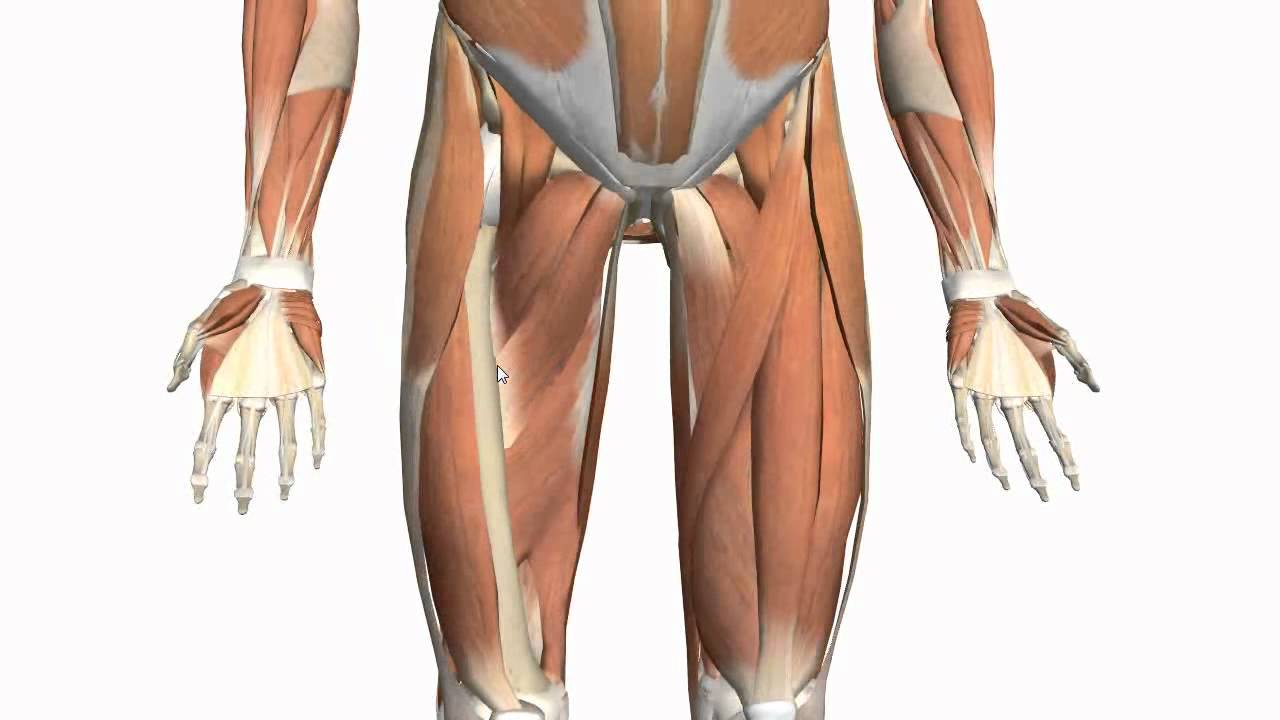
Left Hip Muscles Anatomy : Muscle Anatomy of the Hip. #hipanatomy | Muscle anatomy ... / If you know all the hip flexor names and bones they attach to, that's an awesome accomplishment!
Left Hip Muscles Anatomy : Muscle Anatomy of the Hip. #hipanatomy | Muscle anatomy ... / If you know all the hip flexor names and bones they attach to, that's an awesome accomplishment!. Microscopic anatomy of skeletal muscle. Each muscle below has the bones in bold for intermediate learners and the specific bony landmarks for advanced learners. The hip muscles encompass many muscles of the hip and thigh whose main function is to act on the thigh at the hip joint and stabilize the pelvis. These muscles constitute the anatomical classification known as the medial compartment of the thigh. Learn their anatomy efficiently and easily using kenhub's muscle anatomy and reference charts!
If left unstretched, shortened hip flexors affect the position of the pelvis, which in turn affects the position and movement of the lower back. How muscles are named, 285 hints on how to deduce muscle actions, 286. The cavity of the acetabulum the external obturator muscle is short external rotator muscle of hip joint. The muscular system is responsible for the movement of the human body. Most modern anatomists define 17 of these muscles, although some additional.
Muscles of the Thigh and Gluteal Region - Part 2 - Anatomy ... from i.ytimg.com The muscular system is responsible for the movement of the human body. Meanwhile, labral sulcus and absent labrum are normal variations in the labrum (ring of cartilage). If left unstretched, shortened hip flexors affect the position of the pelvis, which in turn affects the position and movement of the lower back. Several muscles cross the front of the hip and create hip flexion, pulling the thigh and trunk toward each other, but probably the most important is the iliopsoas. Rectus femoris muscle, one of the quadriceps muscles on the front of your thigh. Groin, inguinal region and the anterior. 3 months later i got acute excrutiating pain in inguinal area. Microscopic anatomy of skeletal muscle.
Related online courses on physioplus.
Anterior muscles extend your legs and flex your thighs. Trunk muscles, 289 muscles of the thorax, 289 muscles of the abdominal wall, 289. The main functions of the neck muscles are to permit movements of the neck or head and to provide structural support of the head. The muscles of the pelvis, hip and buttock anatomical chart shows how each muscle in this area of the body works with the others, and the various minor systems within the major ones. Knee assessment and hip mechanics online course: The anterior boundary of the hip adductors is set by if left unchecked, this can lead to chronic knee pain from it band syndrome or acute yet severe injuries such as knee ligament tears (e.g. Anatomy 3d atlas allows you to study human anatomy in an easy and interactive way. Muscle movements, types, and names. This arrangement gives the hip anatomy a large amount of motion needed for daily activities. Major lower body muscle groups include leg and hip muscles, largest muscle groups in your body. The hip muscles encompass many muscles of the hip and thigh whose main function is to act on the thigh at the hip joint and stabilize the pelvis. This webpage presents the anatomical structures found on hip mri. Each muscle below has the bones in bold for intermediate learners and the specific bony landmarks for advanced learners.
I pulled some muscles on left hip hiking. Included within the chart are gorgeous illustrations of the pelvic diaphragm, sphincter muscles, gluteus maximus. Most modern anatomists define 17 of these muscles, although some additional muscles may sometimes be considered. Pelvis and acetabulum, with muscle attachment sites. Rectus femoris forms the middle portion of the quadriceps.
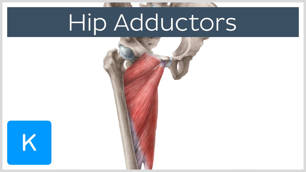
Anatomy of the Hip Adductor Muscles - Human Anatomy ... from i.ytimg.com Each muscle below has the bones in bold for intermediate learners and the specific bony landmarks for advanced learners. Microscopic anatomy of skeletal muscle. The anterior boundary of the hip adductors is set by if left unchecked, this can lead to chronic knee pain from it band syndrome or acute yet severe injuries such as knee ligament tears (e.g. Most modern anatomists define 17 of these muscles, although some additional. Leave a reply cancel reply. The muscular system is responsible for the movement of the human body. If you know all the hip flexor names and bones they attach to, that's an awesome accomplishment! How muscles are named, 285 hints on how to deduce muscle actions, 286.
Most modern anatomists define 17 of these muscles, although some additional muscles may sometimes be considered.
Rectus femoris forms the middle portion of the quadriceps. These muscles constitute the anatomical classification known as the medial compartment of the thigh. 3 months later i got acute excrutiating pain in inguinal area. How muscles are named, 285 hints on how to deduce muscle actions, 286. It is a flat, triangular muscle on the anterior wall of the pelvis. In human anatomy, the muscles of the hip joint are those muscles that cause movement in the hip. Attached to the bones of the skeletal system are about 700 named. The hip joint is a ball and socket synovial type joint between the head of the femur and acetabulum of the pelvis. A bursa that sometimes causes problems in the hip is sandwiched between the bump on the outer hip (the greater trochanter) and the muscles and tendons that cross over the bump. The muscular system is responsible for the movement of the human body. Microscopic anatomy of skeletal muscle. Now that you watched the video, you. Most modern anatomists define 17 of these muscles, although some additional muscles may sometimes be considered.
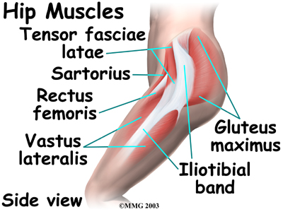
The hip joint is the articulation of the pelvis with the femur, which connects the axial skeleton with the lower extremity. Included within the chart are gorgeous illustrations of the pelvic diaphragm, sphincter muscles, gluteus maximus. It originates at the anterior inferior iliac spine and just above the acetabulum of the hip bone. In human anatomy, the muscles of the hip joint are those muscles that cause movement in the hip. The different anatomical areas of the gluteal region:
Physio Health from 4.bp.blogspot.com Learn about hip muscles human anatomy with free interactive flashcards. The muscles of the pelvis, hip and buttock anatomical chart shows how each muscle in this area of the body works with the others, and the various minor systems within the major ones. Attached to the bones of the skeletal system are about 700 named. The hip muscles encompass many muscles of the hip and thigh whose main function is to act on the thigh at the hip joint and stabilize the pelvis. Anterior muscles extend your legs and flex your thighs. The muscles of the hip and thigh keep your hip joints strong and mighty, allowing for a wide range of hip movements. Related online courses on physioplus. Several muscles cross the front of the hip and create hip flexion, pulling the thigh and trunk toward each other, but probably the most important is the iliopsoas.
The hip flexors are strong, powerful muscles that can overtake the abdominal muscles in some ab exercises.
The hip muscles encompass many muscles of the hip and thigh whose main function is to act on the thigh at the hip joint and stabilize the pelvis. Several muscles cross the front of the hip and create hip flexion, pulling the thigh and trunk toward each other, but probably the most important is the iliopsoas. These muscles constitute the anatomical classification known as the medial compartment of the thigh. In order to isolate the abdominals, you need to minimize the involvement of the hip flexors and maximize the contraction of the abdominals. The main functions of the neck muscles are to permit movements of the neck or head and to provide structural support of the head. Anterior muscles extend your legs and flex your thighs. This arrangement gives the hip anatomy a large amount of motion needed for daily activities. There are a lot of muscles of the hip and thigh. Microscopic anatomy of skeletal muscle. Rectus femoris muscle, one of the quadriceps muscles on the front of your thigh. The muscles of the pelvis, hip and buttock anatomical chart shows how each muscle in this area of the body works with the others, and the various minor systems within the major ones. A bursa that sometimes causes problems in the hip is sandwiched between the bump on the outer hip (the greater trochanter) and the muscles and tendons that cross over the bump. 1 hip anatomy, function and common problems.